26.04.2020
Neighboring neurons' signaling can strengthen or reduce the recipients’ firing rate. They can do this to conserve energy (suppressing over-active, irrelevant, relatively irrelevant signals) and maintain efficient neural processing. This is especially useful for discriminating transient changes in the intensity of visual information (e.g. contours and edges). One direct consequence of this mechanism has been shown with a behavioral experiment in a motion direction discrimination paradigm (Tadin 2003). In this paradigm, observers see a drifting black-white striped grating, the task is to judge the moving direction (e.g. up or down) of the grating. The duration of the stimulus change in each trial depending on the perceptual performance - the better you the individual performs, the lesser (in time) the stimuli stays at the screen. Now, the interesting thing is, the results show that when the grating increases in size, people, on average, needed more time to accurately discriminate the drift direction of the drifting grating. This is very counter-intuitive. You’d think that seeing the thing in a larger size, you would figure out its motion direction easier.
Results show that you need more time to judge the motion direction as accurately as you judge the smaller one’s direction. This is presumed to be driven by the aforementioned mechanism. The neurons encoding the surround send inhibitory signals to the region surrounding the center, thus the performance is suppressed. The image below represents the idea.
This perceptual outcome could be mediated by the neurons encoding the surrounding sending inhibitory signals to the region surrounding the center. The image below represents the idea.
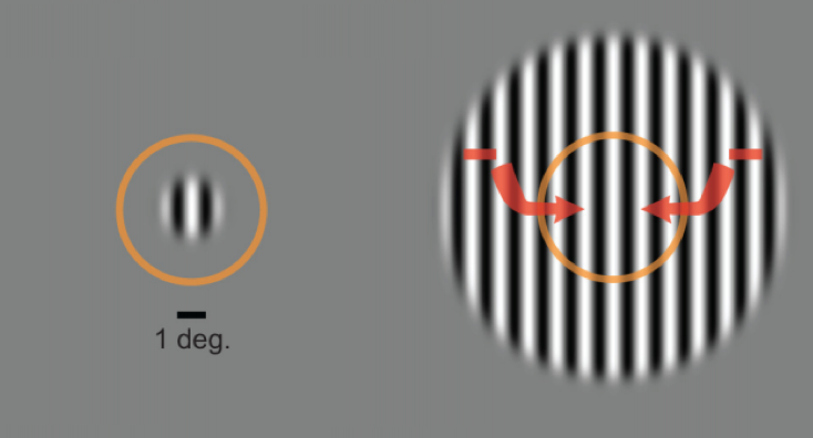
Conversely, and intuitively, when the contrast of the black-white stripes is low, seeing a grating larger helps the performance. This could be due to withdrawal of excitation of the neurons, or increased inhibition, or both.

I’ve joined Huseyin Boyaci and Zahide Pamir to investigate the neural correlates of this behavioral performance at two cortical regions: the primary visual cortex (V1) and the Middle Temporal Area (hMT+). We adopted fMRI to do that. To avoid foveal confluence, we presented the gratings at the periphery of the observer. My main role was to code and analyze the neuroimaging part of the larger experiment.
This is an inflated mesh of both hemispheres of the brain of a participant.
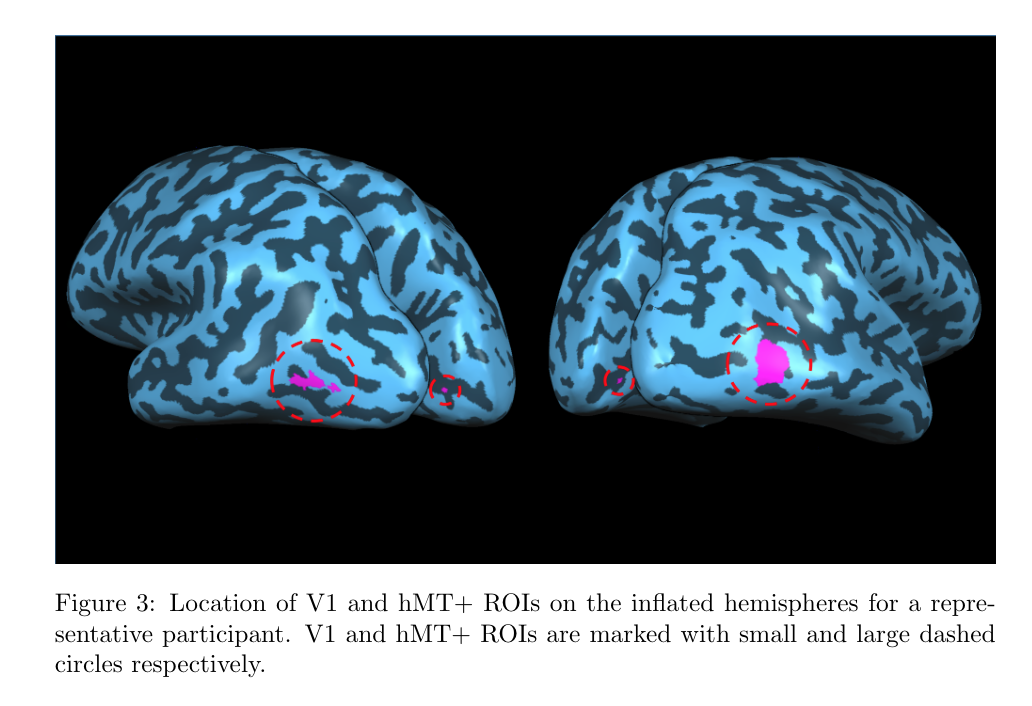
We found that the group of activity of neurons that are responding to the center of the stimuli in the hMT+ region (inferred with BOLD response) appears to be driving the behavioral performance, while the activity of a group of neurons at V1 for the same center of the stimuli does not match with the behavioral outcome. In other words, we found distinct patterns of activity in V1 and hMT+. Here is the link for this project: https://www.sciencedirect.com/science/article/pii/S105381192030570X
More details here in this journal.
Experiment data and codes.
Image credits: Schallmo et al. 2018
References: Tadin, D., Lappin, J. S., Gilroy, L. A., & Blake, R. (2003). Perceptual consequences of centre–surround antagonism in visual motion processing. Nature, 424(6946), 312-315.